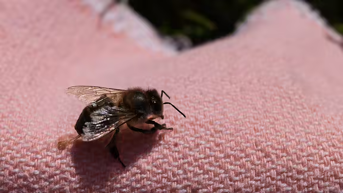
Researchers have directly observed the mechanism that lets a honey bee stinger continue delivering venom after it separates from the insect. High-speed color video showed two flexible valves changing shape as paired lancets move, creating a compact double-piston pump that can operate autonomously inside tissue.
The work, published in the Proceedings of the National Academy of Sciences, addresses a distinctive feature of worker honey bees. A worker can fly away after embedding its stinger, leaving the apparatus to penetrate farther and keep pumping. This self-detachment, known as autotomy, differs from venom systems that rely on muscles surrounding a gland or reservoir.
A honey bee stinger contains three interlocking structural elements that make up its venom canal. A central stylet broadens into the primary pumping chamber. On either side, two sharp lancets slide back and forth to pierce tissue and move venom. Scientists understood those components, but the pumping motion itself had not previously been captured directly.
To study it, the team collected live worker bees, removed their stingers with forceps and placed the stingers partly into artificial gel. Stereomicroscopy and high-speed video recorded the structures in action. Because the components are partly translucent, the researchers could analyze the footage frame by frame and follow the movement inside the chamber.
The recordings showed valves attached to each lancet repeatedly deforming during the cycle. On a downward stroke, a valve expands in a parachute-like shape and seals against the chamber, applying pressure that drives venom toward the target. On the upward movement, it collapses. The valves deform again when they meet near the middle, allowing them to pass each other as the lancets alternate.
That opposed action produces continuing flow while limiting backward movement of venom. Study co-author Fiorella Ramirez-Esquivel of Macquarie University described the valves as collapsible pistons whose geometry allows a small detached mechanism to maintain delivery. Preliminary flow-visualization tests were consistent with what the team saw in the high-speed recordings.
Researchers then used micro-CT scans to build a scaled, 3D-printed model of the pump. Frame-by-frame analysis of that artificial system reproduced the movement seen in biological stingers, strengthening the proposed mechanical explanation.
Questions remain about the valves’ material properties and the possible role of capillary action. The prototype suggested that very high elasticity and low friction are important to the necessary deformation. At the stinger’s scale, capillary forces could help draw venom into the canal or could instead pull fluid from tissue in the opposite direction.
Bee stingers have already influenced microneedles and other tissue-related devices. By explaining how two flexible pistons maintain flow in such a small autonomous package, the new findings may provide another model for miniature medical or microfluidic pumps.